Ergonomics in work place

What is Ergonomics?

Ergonomics is the study of designing and arranging products, systems, and environments in a way that optimizes human well-being, safety, and performance. It aims to reduce discomfort, injuries, and stress while increasing efficiency, productivity, and satisfaction.

Ergonomics is especially crucial in workplaces where individuals perform repetitive tasks or spend long hours in static positions, as it can help prevent musculoskeletal disorders and other work-related injuries. Now we will know about ergonomics in work place.

Office Ergonomics

Office ergonomics is important because it can help prevent work-related injuries and improve overall well-being and productivity of office workers. By designing workstations, chairs, and other equipment in a way that promotes good posture and reduces physical strain, employers can reduce the risk of repetitive stress injuries, back pain, and other musculoskeletal disorders.

 Proper office ergonomics can also help to reduce eye strain, headaches, and other discomfort associated with computer work.

Some common disorders among office employees:

Injury to the muscle and skeletal system is called Musculoskeletal disorders (MSDs). MSDs which occur due to some specific work are called Work related musculoskeletal disorders (WRMSDs).

WRMSDs are common among office employees and can be caused by prolonged sitting or standing, repetitive motions, awkward postures, and insufficient rest breaks. Some of the most common MSDs experienced by office employees include:

  1. Carpal tunnel syndrome
  2. Tendinitis
  3. Trigger finger
  4. Tennis elbow
  5. Rotator cuff injuries
  6. Disc herniation
  7. Repetitive strain injuries (RSIs) etc.

Some common symptoms of above MSDs are:

  1. Neck and shoulder pain: Incorrect posture can cause strain on the neck and shoulders, leading to pain and stiffness.
  2. Back pain: Poor posture can cause back pain, particularly in the lower back, by putting undue stress on the spinal column.
  3. Headaches: Tension in the neck and shoulders can cause tension headaches.
  4. Eye strain: Poor posture can cause eye strain by forcing you to tilt your head or lean forward to see the computer screen.
  5. Tingling and numbness: Poor posture can put pressure on nerves in the arms and legs, leading to tingling and numbness.
  6. Fatigue: Poor posture can lead to fatigue, particularly in the neck and shoulders.

Long-term effects of WRMSDs

If the above symptoms are left untreated the office workers can experience chronic pain, reduced range of motion, decreased muscle strength, and decreased overall quality of life. If left untreated, MSDs can lead to long-term disability, reduced work productivity, and increased healthcare costs.

Repetitive strain injuries (RSIs) such as carpal tunnel syndrome, tendinitis, and tennis elbow can result in chronic pain, reduced grip strength, and reduced hand function. Neck and back pain caused by poor ergonomic practices can lead to chronic pain, limited mobility, and a decreased quality of life.

In addition to the physical effects, long-term MSDs can also lead to psychological stress and mental health issues such as anxiety and depression. This can further impact an employee’s work productivity and quality of life.

Employers can prevent or reduce the long-term effects of MSDs by implementing ergonomic interventions such as providing ergonomic assessments, training on proper posture and stretching techniques, and offering appropriate equipment and workstations. Prompt treatment and rehabilitation of MSDs can also help reduce the risk of long-term effects.

Benefits of  following ergonomics:

  1. Increased comfort: Ergonomic design can help reduce discomfort, fatigue, and pain associated with prolonged sitting or standing.
  2. Improved productivity: If employees follow ergonomics it can improve work efficiency, accuracy, and speed. 
  3. Reduced injury risk: Ergonomic design can reduce the risk of musculoskeletal disorders and other workplace injuries.
  4. Better posture: Ergonomic design can help promote good posture, which can reduce the risk of neck and back pain.
  5. Improved employee morale: Providing an ergonomic workspace can improve employee satisfaction, leading to higher morale and job satisfaction.
  6. Cost savings: Implementing ergonomic design can lead to cost savings by reducing absenteeism, turnover, and workers compensation claims.

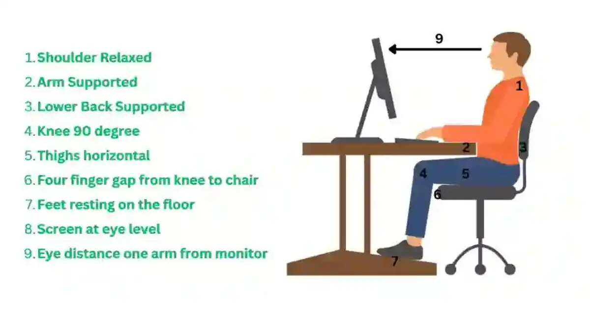
Here are some tips for an ergonomic office setup:

  1. Chair: Use a chair with adjustable height, backrest, and armrests. Ensure that your feet are flat on the floor and your hips are level with or higher than your knees. Adjust the backrest to support your lower back.
  2. Desk: Use a desk with adjustable height that allows your forearms to rest comfortably on the desk and your wrists to be straight while typing. Position your computer monitor at eye level, an arm’s length away from your body.
  3. Keyboard and Mouse: Use an ergonomic keyboard and mouse to reduce strain on your hands and wrists. Keep the keyboard and mouse at the same level and close to your body to avoid reaching and straining.
  4. Lighting: Use proper lighting to avoid eye strain and reduce glare on the computer screen. Use task lighting to illuminate specific work areas.
  5. Posture: Sit up straight with your shoulders relaxed and your head level. Avoid slouching or leaning forward.
  6. Breaks: Take frequent breaks to stand up, stretch, and move around. This can help reduce the risk of fatigue and strain.
Ergonomics office setup

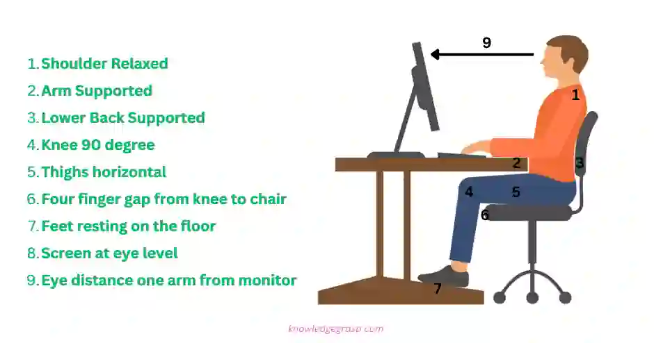
By setting up your office ergonomically, you can help reduce the risk of musculoskeletal disorders (MSDs) and improve your work efficiency and productivity.

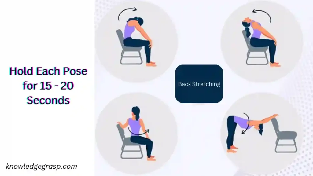
Stretching exercises in work place

Stretching exercises can be helpful in reducing muscle tension, increasing blood flow, and improving flexibility, particularly for individuals who spend extended periods sitting in front of a computer. Here are some stretching exercises that can be done in an office setting:

  1. Neck stretch: Gently tilt your head to one side, bringing your ear toward your shoulder. Hold for 10-15 seconds and repeat on the other side.
  2. Shoulder roll: Roll your shoulders forward and backward in a circular motion for 10-15 repetitions.
  3. Seated spinal twist: While seated, cross one leg over the other and twist your torso toward the leg that is crossed. Place your opposite hand on the outer thigh and hold for 10-15 seconds. Repeat on the other side.
  4. Hamstring stretch: While seated, extend one leg straight out and reach for your toes. Hold for 10-15 seconds and repeat on the other side.
  5. Wrist stretch: Hold your arm out in front of you with your palm facing down. Use your opposite hand to gently pull your fingers back toward your wrist. Hold for 10-15 seconds and repeat on the other side.
Office stretching exercise

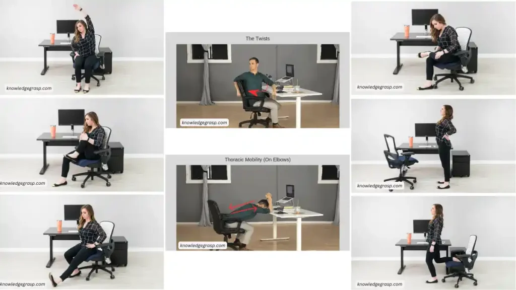
Here are some stretching exercises for low back pain that can be done in the office:

  1. Seated forward fold: Sit in your chair with your feet flat on the floor. Slowly fold forward from your hips, reaching toward your feet or the floor. Hold for 10-15 seconds and slowly return to a seated position.
  2. Figure four stretch: Sit in your chair and cross one ankle over the opposite knee. Gently press down on the crossed leg until you feel a stretch in your buttocks and lower back. Hold for 10-15 seconds and repeat on the other side.
  3. Seated twist: Sit in your chair with your feet flat on the floor. Twist your torso to one side, holding the back of your chair for support. Hold for 10-15 seconds and repeat on the other side.
  4. Cat-cow stretch: Sit on the edge of your chair with your feet flat on the floor. Place your hands on your thighs and arch your back, tucking your chin into your chest. Then slowly round your back, dropping your head forward. Repeat for 10-15 repetitions.
  5. Hip flexor stretch: Sit on the edge of your chair with one knee bent and the other leg extended behind you. Gently press your hips forward until you feel a stretch in your hip flexor. Hold for 10-15 seconds and repeat on the other side.
  6. Back extension: Stand in your place and keep both hands on your lower back for support. Lean backward and hold the position for 15-20 seconds and repeat every one hour.
Back stretching in office

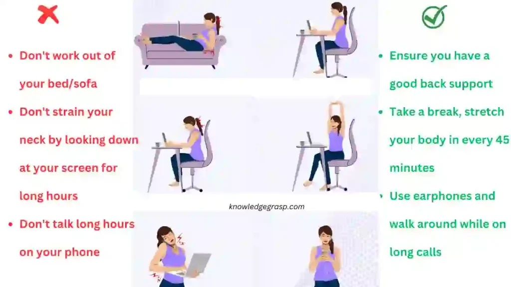
Ergonomics in work place( Do’s & Don’ts )

do's & don'ts of ergonomics

Exercise-induced Muscle Soreness or Delayed Onset Muscle Soreness (DOMS)

Exercise Induced Muscle Soreness

Exercise induced muscle soreness, also known as delayed onset muscle soreness (DOMS) or “muscle fever”, is a common experience for many people who engage in physical activity. It is the pain and stiffness felt in muscles several hours to days after an unusual or strenuous physical activity. The intensity of muscle soreness can vary from person to person and can range from mild discomfort to severe pain. It usually peaks 24 to 48 hours after the activity and can last for several days.

More about delayed onset muscle soreness (DOMS)

The cause of Exercise Induced Muscle Soreness

It is believed to be due to microscopic tears in the muscle fibers and the inflammation that occurs in response to the injury. This type of muscle damage occurs when the muscle is subjected to eccentric contractions, which occur when the muscle is lengthening while under tension. Examples of eccentric contractions include the lowering phase of a bicep curl or the landing phase of a jump.

Risk factors for developing DOMS

It include performing new or unfamiliar exercises, increasing the intensity or duration of a workout too quickly, and poor conditioning. DOMS is more likely to occur in individuals who are new to exercise or have been inactive for a prolonged period of time. However, even well-trained athletes can experience DOMS if they significantly increase the intensity or volume of their training.

Symptoms of DOMS

It can include muscle pain and stiffness, weakness, decreased range of motion, and swelling. The affected muscle may also feel tender to the touch and may be sore to the point of feeling bruised. These symptoms usually peak 24 to 48 hours after the exercise and can last for several days.

To reduce muscle soreness, people often use rest, ice, gentle stretching, and over-the-counter pain relievers. It’s also important to stay hydrated and get enough protein. Gentle exercise, such as a light jog or bike ride, can also help to alleviate muscle soreness by increasing blood flow to the affected area.

In physical therapy, delayed onset muscle soreness (DOMS) is a common issue that can be addressed through various treatments and interventions. Physical therapists use a variety of techniques to help alleviate muscle soreness, including:

  1. Stretching: Gentle stretching can help to increase range of motion and decrease muscle soreness. Physical therapists will typically recommend specific stretches for the affected muscle or muscle group.
  2. Massage: Massage therapy can help to increase blood flow and decrease muscle soreness. Physical therapists may use techniques such as soft tissue mobilization or myofascial release to help alleviate muscle soreness.
  3. Ice or Cold Therapy: Cold therapy can help to reduce inflammation and decrease muscle soreness. Physical therapists may use ice packs or cryotherapy to help alleviate muscle soreness.
  4. Heat therapy: Heat therapy can help to increase blood flow and decrease muscle soreness. Physical therapists may use heat packs or therapeutic ultrasound to help alleviate muscle soreness.
  5. exercises: physical therapists may use different exercises to help alleviate muscle soreness. These exercises may include range of motion exercises, strengthening exercises, and endurance exercises.
  6. Education: physical therapists may provide education to the patient on how to properly warm up and cool down before and after exercises, how to adjust the intensity of their workout, and how to properly perform exercises to prevent DOMS and future injuries.

Physical therapists work with patients to develop an individualized treatment plan that addresses their specific needs and goals. They may also work with patients to improve their overall fitness and prevent future episodes of DOMS.

It’s important to note that while muscle soreness is a normal part of exercise, it should not be ignored. If the pain is severe or persists for more than a few days, it is important to seek physical therapy attention. In some cases, muscle soreness may be a sign of a more serious injury, such as a muscle strain or tear, and a physical therapist can help determine the cause of the pain and develop an appropriate treatment plan.

In conclusion, muscle soreness is a common experience for many people who engage in physical activity. It is caused by microscopic tears in the muscle fibers and the inflammation that occurs in response to the injury. The intensity of muscle soreness can vary from person to person and can range from mild discomfort to severe pain. It usually peaks 24 to 48 hours after the activity and can last for several days. To reduce muscle soreness, people often use rest, ice, gentle stretching, and over-the-counter pain relievers. It’s also important to stay hydrated and get enough protein. Gentle exercise, such as a light jog or bike ride, can also help to alleviate muscle soreness by increasing blood flow to the affected area.

Continuous Passive Movement (CPM)

Continuous passive movement (CPM)

Continuous passive movement (CPM) is a type of therapy that involves using a machine to move a joint through a range of motion without the patient actively participating. It is typically used to improve range of motion and reduce pain and stiffness in joints that have been immobilized, such as after surgery or an injury.

CPM is typically used for the knee, ankle, elbow, or shoulder joint. The machine is adjusted to the patient’s range of motion and then moves the joint through a set range of motion at a slow and steady pace. The patient sits or lies down and the machine is attached to the affected joint.

The CPM machine is designed to move the joint at a slow, consistent pace, typically between 30 and 90 degrees per minute. The therapy is typically administered for a period of 30 minutes to 2 hours per day, depending on the patient’s condition and the physician’s recommendations. The therapy is typically started within 24-48 hours of surgery and can last for up to 3-6 weeks post-surgery.

Types of Continuous Passive Movement (CPM)

There are different types of CPM machines available, including those designed for the knee, hip, ankle, elbow, and shoulder. The machines vary in size, weight, and cost, and can be either portable or stationary. Some CPM machines can be used at home, while others are only available in a clinical setting.

In addition, there are also some other variations of CPM machines that are used to target different areas of the body or to provide different types of therapy. Some examples include:

  • Multi-joint CPM: This type of machine is designed to move multiple joints in the body at the same time. It can be used to treat patients with multiple joint injuries or surgeries.
  • Spinal CPM: This type of machine is designed to move the spine through a range of motion. It is often used to help patients with spinal conditions such as scoliosis or spinal fractures regain range of motion and reduce pain.
  • Hand and Wrist CPM: This type of machine is designed to move the hand and wrist through a range of motion. It is often used to help patients with hand and wrist injuries or surgeries regain range of motion and strength.
  • Pediatric CPM: This type of machine is designed for use in children. It can be used to help children with injuries or surgeries regain range of motion and reduce pain.
  • Dynamic CPM: This type of machine is designed to provide a more active form of therapy by using a combination of passive and active movements. It is often used to help patients with more severe injuries or surgeries regain range of motion and reduce pain.

The therapy is typically performed for several hours a day, for several weeks. It is typically used in the early stages of rehabilitation, as a way to maintain and improve range of motion while the patient is still unable to actively move the joint.

Indications of CPM

Continuous Passive Movement (CPM) therapy is typically used to treat a variety of conditions related to joint injury or surgery. Some common indications for CPM therapy include:

  • Knee replacement surgery: CPM therapy is often used to help patients regain range of motion and strength in the knee following surgery.
  • Hip replacement surgery: Similar to knee replacement, CPM therapy can be used to help patients regain range of motion and strength in the hip following surgery.
  • Traumatic joint injuries: CPM therapy can be used to help patients regain range of motion and reduce pain following a traumatic injury to a joint such as a fracture, dislocation, or ligament tear.
  • Soft tissue injuries: CPM therapy can be used to help patients regain range of motion and reduce pain following injuries to the tendons, ligaments, or muscles surrounding a joint.
  • Arthritis: CPM therapy may be used to help patients with arthritis maintain or improve range of motion in the affected joint.
  • Rehabilitation after cast immobilization: CPM therapy can be used to help patients regain range of motion following a period of immobilization in a cast.

It’s important to note that CPM therapy is typically used as part of a comprehensive treatment plan, and is often used in conjunction with other forms of therapy such as exercise and manual therapy. A Physical Therapist or physician will determine if CPM is suitable for the patient and set the appropriate course of treatment.

Contraindications of CPM

Continuous Passive Movement (CPM) therapy is generally considered safe, but there are certain contraindications that should be considered before starting the therapy. These include:

  • Deep vein thrombosis: CPM therapy should not be used on patients who have a history of deep vein thrombosis, as the therapy may increase the risk of blood clots.
  • Bone infection: CPM therapy should not be used on patients who have an active bone infection, as the therapy may spread the infection.
  • Vascular insufficiency: CPM therapy should not be used on patients who have poor circulation in the affected limb, as the therapy may increase the risk of tissue damage.
  • Sensory or motor deficits: CPM therapy should not be used on patients who have significant sensory or motor deficits in the affected limb, as the therapy may not be effective or may cause further damage.
  • Open wounds or skin breakdown: CPM therapy should not be used on patients who have open wounds or skin breakdown in the area of the joint, as the therapy may cause further irritation or damage.
  • Metal allergies: CPM therapy should not be used on patients who have metal allergies, as the therapy may cause adverse reactions.

It’s important to note that CPM therapy should be used under the guidance of a physical therapist or physician, as they will evaluate the patient’s condition and determine if CPM therapy is appropriate. If CPM is contraindicated, alternative therapies may be recommended.

Overall, continuous passive movement therapy is a non-invasive and safe way to help improve range of motion, reduce pain and stiffness, and prevent muscle atrophy. It should be used under the guidance of a physical therapist or other healthcare professional, and it may not be suitable for everyone.

Nerve Conduction Velocity (NCV)

Nerve conduction velocity (NCV) is a measure of the speed at which electrical impulses travel through a nerve. It is typically measured in meters per second (m/s) or feet per second (ft/s). The NCV of a nerve can be affected by a number of factors, including the thickness of the nerve fibers, the degree of myelination, and the presence of any structural or metabolic abnormalities. NCV is commonly used in the diagnosis of various neurological conditions, such as peripheral neuropathy, carpal tunnel syndrome, and Guillain-Barré syndrome. It is typically measured using a nerve conduction study (NCS), which involves the application of small electrical stimuli to a nerve and the recording of the resulting muscle or nerve responses.

Types of nerve conduction velocity (NCV) tests:

  1. Motor nerve conduction study (MNCS)
  2. Sensory nerve conduction study (SNCS)

Both types of NCV tests are non-invasive, painless and usually performed as an outpatient procedure. The results of the test are analyzed and interpreted by a neurologist who can diagnose underlying neurological conditions if present.

Motor nerve conduction velocity (NCV) test is a diagnostic tool that measures the speed of electrical impulses as they travel through a motor nerve. Motor nerves are responsible for controlling muscle movement. During the test, small electrodes are placed on the skin over the nerve being tested, and a mild electrical current is applied. The electrical activity of the nerve is then measured and recorded. The test typically measures the conduction velocity of the nerve, which is the speed at which the electrical impulse travels along the nerve.

The results of the test are compared to normal values to determine if there is any nerve damage or dysfunction. Motor NCV test is often used to diagnose and evaluate nerve damage or disorders such as peripheral neuropathy, carpal tunnel syndrome, and Guillain-Barre syndrome. It is commonly used in combination with sensory NCV test to have a complete picture of the nerve conduction.

During the test, the electrode at the muscle will pick up the muscle’s response to the electrical stimulus and this response is called the compound muscle action potential (CMAP) which will give the amplitude, latency and conduction velocity of the motor nerve. These values will be compared with normal values to see if there is any damage or dysfunction in the motor nerve.

A sensory nerve conduction velocity (NCV) test is a test that measures the speed at which electrical impulses travel through a sensory nerve. Sensory nerves are responsible for carrying information about touch, temperature, pain, and other sensations from the skin and other tissues to the brain.

During the test, small electrodes are placed on the skin over the nerve being tested, and a mild electrical current is applied. The electrical activity of the nerve is then measured and recorded. The test typically measures the conduction velocity of the nerve, which is the speed at which the electrical impulse travels along the nerve. The results are compared to normal values to determine if there is any nerve damage or dysfunction.

Sensory NCV tests are also often used to diagnose and evaluate nerve damage or disorders  peripheral neuropathy, carpal tunnel syndrome, and Guillain-Barre syndrome. It is commonly used in combination with motor NCV test to have a complete picture of the nerve conduction.

Principles of nerve conduction study:

The principles of a nerve conduction study (NCS) involve measuring the electrical activity of a nerve as it conducts an impulse. The test uses small electrodes placed on the skin over the nerve being studied, and a mild electrical current is applied to the nerve. The electrical activity of the nerve is then measured and recorded, which provides information about the health and function of the nerve.

The main principles of NCS are:

  1. Excitability: This principle refers to the ability of a nerve to respond to a stimulus. In NCS, the nerve’s excitability is measured by the amplitude of the electrical response.
  2. Conduction velocity: This principle refers to the speed at which an impulse travels through a nerve. In NCS, the nerve’s conduction velocity is measured by the time it takes for the impulse to travel from one electrode to another.
  3. Latency: This principle refers to the time it takes for a nerve to respond to a stimulus. In NCS, the nerve’s latency is measured by the time between the application of the electrical stimulus and the onset of the electrical response.
  4. Distance: The distance of the nerve is a important parameter in NCS, the long the distance of the nerve, the longer the latency will be.

By measuring these parameters, NCS can provide valuable information about the health and function of a nerve, and can be used to diagnose and evaluate nerve damage or disorders.

Variables/factors affecting NCV study:

Nerve conduction velocity (NCV) is the speed at which electrical impulses travel through a nerve. The main physiological variables that affect NCV include:

  1. Axon diameter: Larger axons have a faster NCV compared to smaller axons.
  2. Myelination: Myelinated nerves have a faster NCV than unmyelinated nerves.
  3. Temperature: Increased temperature can lead to faster NCV, while decreased temperature can slow it down.
  4. Nerve fiber type: Different types of nerve fibers (e.g. A-beta, A-delta, and C fibers) have different NCV.
  5. Age: NCV tends to decrease with age.
  6. Sex: NCV may vary between males and females.
  7. Disease or injury: Certain diseases or injuries (e.g. diabetes, peripheral neuropathy) can affect NCV.
  8. Drugs and toxins: Some drugs and toxins can alter NCV.
  9. Hormonal status: Hormonal status can affect NCV.
  10. Metabolic state: metabolic state can affect NCV.

In addition to the physiological variables that affect nerve conduction velocity (NCV), there are also technical variables that can impact the measurement of NCV. These include:

  1. Stimulus intensity: The strength of the electrical stimulus used to elicit a nerve response can affect the measured NCV.
  2. Distance between electrodes: The distance between the stimulating and recording electrodes can affect the measured NCV.
  3. Electrode size: The size of the electrodes used to stimulate and record the nerve response can affect the measured NCV.
  4. Electrode placement: The location of the electrodes on the skin can affect the measured NCV.
  5. Filtering settings: The settings used to filter the electrical signals can affect the measured NCV.
  6. Amplitude and duration of the stimulus: The amplitude and duration of the stimulus can affect the measured NCV.
  7. Temperature and humidity of the environment: Temperature and humidity of the environment can affect the measured NCV.
  8. The quality of the equipment: The quality of the equipment used to measure NCV can affect the accuracy of the measurement.
  9. The technique used: The technique used to measure the NCV such as motor or sensory can affect the measurement.
  10. The measurement protocol: The specific protocol used to measure the NCV can affect the measurement.

It is important to control for these technical variables and use appropriate techniques to ensure accurate and reliable measurement of NCV.